ATF: Biomed ENG
Takahashi Lab


ATF: Biomed ENG
Takahashi Lab


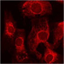
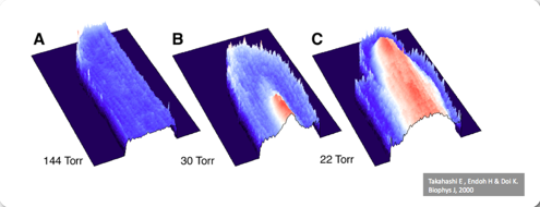
We are developing novel techniques for imaging oxygen transport to and within cells. Using these techniques, we have demonstrated mechanisms by which cells undergo necrotic cell death even at physiological oxygen concentrations.
Follow this link for more.
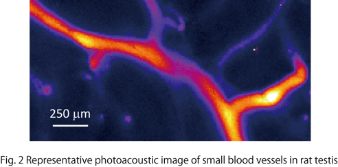
•Photoacoustic imaging to visualize deep structures in living tissues (Yamaoka)
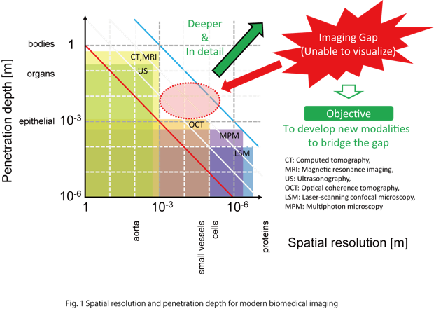
Accurate detection of the growth extent of cancer cells in depth direction is very important. In particular, depth discrimination of melanoma cells is extremely essential because even a tiny difference in the invasion of cancer cells strongly determines the outcome for the patients . Depth resolution less than 10 µm (size of cells) is required to discriminate individual cells. As for the imaging depth, several mm is required, because the thickness of skin tissues is of several-mm order. Imaging with 10-µm resolution is also important to visualize small vascular channels, key constituents in living tissues. In this way, imaging over several-mm depth with 10-µm spatial resolution is desired in clinical applications.
Optical imaging typified by laser-scanning microscopy has a spatial resolution high enough to observe single cells. However the penetration depth of more than 1 mm is difficult to obtain. On the other hand, magnetic resonance imaging (MRI), computed tomography (CT) and ultrasonography (US), which are commonly used in medicine, can visualize deeply enough to investigate whole human bodies. However, the spatial resolution is not high enough to observe single cells. Thus, imaging modalities with a spatial resolution of 10 µm at around several mm depth do not exist (Fig. 1).
To bridge this gap in imaging modalities, we have studied and developed various types of photoacoustic imaging (especially, two-photon photoacoustic microscopy (TP-PAM, Fig. 2).
•A life with less oxygen? (Takahashi)
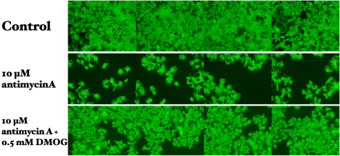
Recently, we have demonstrated that some cancer cell lines can sustain mitochondrial membrane potential without oxygen when cellular oxygen sensor (PHD/HIF-1) is activated.
Takahashi E and Sato M: Anaerobic respiration sustains mitochondrial membrane potential in prolyl hydroxylase pathway-activated cancer cell line in a hypoxic microenvironment. Am J Physiol-Cell Physiol (accepted for publication on Sep. 11, 2013)
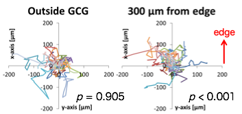
To elucidate the initial mechanisms of hematogenous metastasis of cancer cells, we hypothesized that cancer cell might migrate towards regions with higher oxygen concentration such as intratumor micro vessels along the gradient of oxygen concentration. To test this hypothesis, we have devised the gap cover glass (GCG) by which oxygen concentration gradients were established in monolayer cultured cells. We demonstrated a directional migration of MDA-MB-231 cells under the oxygen gradient.
•Development of compound sensors and multi-imaging systems (Kimoto)
This research project includes development of sensing systems including;
•Multi-modality imaging system
•Multifunctional object recognition system
•Simultaneous measurement of EMG/MMG/NIRS for applications in medicine
•Skin and hair condition measurements
•Environment measurement systems


•Bioimaging of cellular oxygen transport and metabolism (Takahashi)
•Cancer cells prefer higher oxygen? (Takahashi)